Medtronic spacer
Medtronic
Medtronic
Medtronic Home > Information for Physicians > Pain > Pain Therapies > Diagnosis
  Pain Therapies
 

Introduction to Chronic Pain


Types of Pain


Chronic Back & Leg Pain


Complex Regional Pain Syndromes


Cancer Pain


Diagnosis


What is Neurostimulation ?


What is Intrathecal Drug Delivery ?

Medtronic
Medtronic

Medtronic
Medtronic

Diagnosis

In this section you can find information about the diagnosis of Failed Back Syndrome and of Complex Regional Pain Syndrome.

Causes and symptoms of FBS

FBS can be caused by: [1-7]
 
  • irreversible nerve injury
  • surgical complications
  • psychosocial problems
  • inappropriate selection of patients for surgery
  • recurrent herniation
  • wrong level of operation
Patients with FBS may suffer from: [3,4,8]
 
  • lumbosacral postoperative fibrosis and/or arachnoiditis
  • root lesion
  • dorsal compartment syndrome
  • lateral spinal stenosis

Chronic back and leg pain associated with FBS

Most patients with FBS suffer from mixed back and leg pain, which is often of a
combined neuropathic and nociceptive nature.

FBS always involves the dorsal horn, having central sensitisation and mostly
sympathetic involvement.


FBS consisting primarily of leg pain includes:
 
  • A mixture of nociceptive and neuropathic pain or
  • Neuropathic pain essentially (or primarily) caused by:
    • nerve damage (pre-operation, post-operation and wrong level of operation); epidural fibrosis;
    • lateral spinal stenosis
FBS consisting primarily of back pain is caused by: degeneration of intervertebral joints; pseudoarthrosis, an infection or a degenerative change at other vertebrae; lumbar facet
syndrome; facet joints disc degeneration; lateral spinal stenosis; spondylitis; problems
on another level.

Diagnosis of common conditions leading to low back and/or leg pain

Source of pain

Key characteristics

Clinical examination
Other examinations
Disc degeneration

1. Predominantly  located in the low back area, may radiate out to gluteal zone(s)

2. May radiate to lower limb(s), in a non-dermatome distribution

3. Increases during activities that increase strain on the disc, such as sitting, bending or lifting weights

4. Improves when  resting or walking around

1. In most cases lumbar flexion is restricted and painful

2. Usually low-lumbar pressure sensitivity

3. No neurological abnormalities or signs of root compression
1. X-rays usually show narrowing of one or  more intervertebral spaces; osteophyte growth may be present

2. MRI scans usually show dehydration of  the disc
Disc herniation 1. Pain is mostly dermatome-bound and course corresponds to the spinal nerve innervation

2. Sensory disorders in the affected dermatome may be described

3. There may be a reduction in the strength of the muscles associated with the affected nerve
1. Demonstrates neurological abnormalities, such
as sensory disorders, weakened reflexes
and motor weakness


2. The Lasègue test or femoralis test is usually positive
1. Hernia can be seen on a CT or MRI scan

2. EMG and meticulous neurological examination confirms which root is compressed
Facet joint pain Pain usually lateral to the midline, often accompanied by discogenic back pain 1. Signs not specific

2. Patients usually feel pain on extension

3. Often paravertebral elective pressure pain in the facet joints
1. Radiological images, particularly the ¾-rotation image, can show the facet joint compression

2. CT scans can show narrowed and hypertrophic facet joints

3. Infiltration of the joint with local anaesthetic and a corticoid preparation, under fluoroscopy or CT support, results in immediate pain relief
Spinal stenosis 1. Signs not specific

2. Patients usually feel pain on extension

3. Often paravertebral elective pressure pain in the facet joints
1. Signs not specific

2. Medical history usually allows a diagnosis to be made
1. Diagnosis suspected if the radiological images show a degenerative antero-/laterolisthesis

2. Diagnosis can be confirmed by a CT scan, MRI or myelography

References
1. Anderson VC et al. Current review of pain 2000;4:105-11
2. Leveque JC et al. Neuromodulation 2001;4:1-9
3. Dario A et al. Neuromodulation 2001;4:105-110
4. Ohnmeiss DD et al. The Spine Journal 2001:358-363
5. Vaccaro AR et al. Spine 2001;26 (24): S111-8
6. Spengler DM et al. Spine 1980;5:356-60
7. Fager CA, Freidberg SR. Spine 1980;5:87-94
8. Long DM. Surgical management of pain 2002: 354-64


Signs and symptoms of CRPS

Sensory
  • Intense pain [1,2]
  • Hyperesthesia [1,3]
  • Hyperalgesia [1,3]
  • Allodynia [1,3]
  • Sensory deficits e.g. hemisensory impairment [4]
  • Trigeminal hypoesthesia [5]
Autonomic
  • Swelling [1]
  • Colour and temperature changes [6]
  • Sweating abnormalities [6]
Motor dysfunction
  • Weakness
  • Decreased range of motion [3]
  • Tremor [1,6]
  • Dystonia [7]
  • Myoclonus [7]
  • Decreased muscular strength, especially grip strength [8]
Dystrophic
  • Increased or decreased nail and hair growth
  • Skin changes e.g. palor, plantar fibrosis, hyperkeratosis and thin glossy skin [9]

Intense pain and hyperesthesia are the predominant sensory symptoms. The pain, characterized as aching, burning, pricking or shooting, is localized deep in the somatic tissue.

Sensory deficits are common. Rommel et al. observed that 33% of patients had hemisensory impairment with decreased temperature and pinprick sensation ipsilateral to the affected limb. [4]

Trigeminal hypoesthesia has been found in 49% of CRPS patients with upper extremity disease versus <10% in patients with other pain and normal individuals. [5]

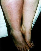
The majority of patients describe swelling of the affected limb, which can be aggregated by physical load, painful stimuli and environmental and local temperature changes.

Temperature asymmetry between the affected and unaffected side measured with infrared thermography exceeds 1°C. [3] Such asymmetry can be warmer or colder.

Weakness, tremor and reduced movement are frequently seen.

Zyluk observed that 78% of patients had significantly reduced grip strength. [8]

Range of motion is decreased by joint effusion early in the disease and by contraction and
fibrosis later in the disease. [3]

Tremor has been reported in 24% to 60% of patients. [6,10]

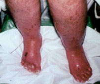
Although dystrophic changes are generally described as occurring late in the disorder, they may appear within weeks of its onset. In some cases, allodynia may be so severe that the extremity is held in a protective posture further accelerating the development of dystrophic changes in both integument and deeper structure


Courtesy of Robert J. Schwartzman, M.D. MCP Hahnemann School of Medicine

International Association for the Study of Pain (IASP) Diagnostic

Type I (Reflex Sympathetic Dystrophy)
Type II (Causalgia)
1. The presence of an initiating noxious event, or a cause of immobilization 1. The presence of continuing pain, allodynia, or hyperalgesia after a
nerve injury, not necessarily limited to
the distribution of the injured nerve
2. Continuing pain, allodynia, or hyperalgesia with which the pain is disproportionate to any inciting event 2. Evidence of oedema, changes in skin blood flow, or abnormal sudomotor activity in the region of the pain
3. Evidence of oedema, changes in skin blood flow, or abnormal sudomotor activity in the region of the pain 3. This diagnosis is excluded by the existence of conditions that would otherwise account for the degree of pain and dysfunction
4. This diagnosis is excluded by the existence of conditions that would otherwise account for the degree of pain and dysfunction  
Criteria 2-4 must be satisfied
All three criteria must be satisfied

References
1. Harden RN et al. Pain 1999;83:211-219
2. Galer BS et al. J Pain Symptom Manage 2000;20:286-92
3. Birklein F, Handwerker HO. Pain 2001; 94:1-6
4. Rommel O et al Pain 1999;80:95-101
5. Thimineur M et al. Clin J Pain 1998;14:256-67
6. Birklein F et al. Pain 1997;69:49-54
7. Schwartzman RJ, Kerrigan J. Neurosurgery 1990;40:57-61
8. Zyluk A. J Hand Surg 2001;26:151-154
9. Wasner G et al. Neuro Clin 1998;16:851-68
10. Sieweke N et al. Pain 1999;80:171-177
11. Stanton-Hicks MD et al. Clin J Pain 1998;15:155-166
12. Merskey H, Bogduk H. IASP Press 1994; pp40-43.


back
 

top
 

Privacy Statement Terms of Use Medtronic Footer